BIOS 242 Week 3 Lab 1; Differential Staining
-
$20.00
| Institution | BIOS 242 Fundamentals of Microbiology with Lab - Chamberlain |
| Contributor | Anika Fultz |
Lab 5: Differential Staining
Learning Objectives:
- Explain the principle of differential staining
- Apply differential staining to identify bacteria
Differential stain uses two or more stains to specifically stain certain structures or cellular components which cannot be easily observed using simple stains. Differential staining principles are based upon the specific chemical nature and composition of cellular components and therefore, different structures are observed using different stains and staining procedures. Differential staining often becomes the basis of identification of the bacteria in clinical labs.
In this lab, we will learn about four separate staining procedures.
- Gram Staining
- Endospore Staining
- Acid-fast Staining
- Capsule Staining (Special Staining)
Exercise 1: Gram Staining
Materials:
Broth cultures of S. epidermidis, E. coli, a mixed culture containing one Gram positive and one Gram negative bacteria; sterile loop, Gram staining kit, glass slides, Incinerator, DI water, marker, immersion oil, lens paper, bibulous paper, microscope
Method:
- Obtain glass slides and pure cultures. Write the name of the bacteria on the side of the glass slide.
- Using aseptic technique make a thin smear. Allow it to air dry and then heat fix it.
- Add a drop or two of crystal violet and allow staining for 1 minute.
- Gently wash off crystal violet using a few drops of DI water.
- Add a few drops of iodine solution to the slide and wait for 1 minute.
- Wash iodine solution using a few drops of water......... Continue
Exercise 2: Differential Staining- Observation of prepared slides
Materials:
Prepared slides of endospore, capsule and acid-fast staining; immersion oil, lens paper, microscope
Method:
- Obtain the prepared slides.
- Observe slides using the 10X, and 40X objective lenses.
- Record observations made using 40X objective lens in the lab report.
- Complete the lab report.
Lab Report:
Purpose: Describe the purpose of the lab.
Exercise 1: Gram Staining
Draw the observation inside the circles provided. Label your image appropriately. Write the magnification at which the observation was made.
S. epidermidis:
Additional observation/notes:
E. coli:
Additional observation/notes:
Mixed culture:
Additional observation/notes:
Questions:
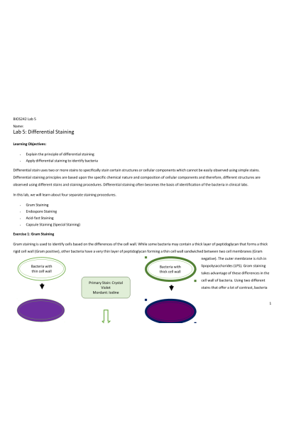
Complete the following table by predicting colors of bacteria with- and thin cell walls as they are processed through the steps of Gram staining.
- A fellow student showed you a Gram stained slide where cells containing thick cell walls were stained pink. What would you tell her about the staining procedure? Why?
- A fellow student showed you a Gram stained slide where cells containing LPS and a thin peptidoglycan layer were stained purple. What would you tell her about the staining procedure? Why?
Exercise 2: Differential Staining- Observation of prepared slides
Draw the observation inside the circles provided. Label your image appropriately. Write the magnification at which the observation was made.
Endospore staining:
Acid-Fast Staining:
Additional observation/notes:
Capsule staining:
Questions:
- Describe the differences between simple staining and differential staining principles.
- A student forgot to use heat during the endospore staining procedure. How will it impact the observation?
- Why is the capsule staining called a negative staining method?
- What is meant by the term “acid-fast”?
| Instituition / Term | |
| Term | Uploaded 2023 |
| Institution | BIOS 242 Fundamentals of Microbiology with Lab - Chamberlain |
| Contributor | Anika Fultz |


