BIOS 242 Week 2 Assignment; Lab 4 of 14 Onsite; Simple Staining
-
$20.00
| Institution | Chamberlain |
| Contributor | Nikki |
Document Preview
Lab 4: Simple Staining
Learning Objectives:
- To learn about principle of staining
- To learn the process of simple stain
- To prepare wet mount slides
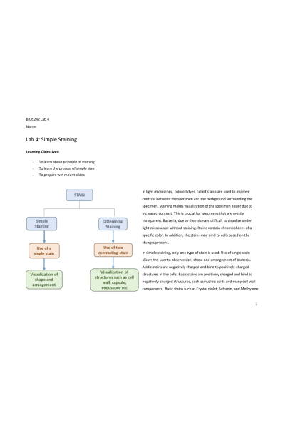
In light microscopy, colored dyes, called stains are used to improve contrast between the specimen and the background surrounding the specimen. Staining makes visualization of the specimen easier due to increased contrast. This is crucial for specimens that are mostly transparent. Bacteria, due to their size are difficult to visualize under light microscope without staining. Stains contain chromophores of a specific color. In addition, the stains may bind to cells based on the charges present. In simple staining, only one type of stain is used. Use of single stain allows the user to observe size, shape and arrangement of bacteria. Acidic stains are negatively charged and bind to positively-charged structures in the cells. Basic stains are positively charged and bind to negatively-charged structures, such as nucleic acids and many cell wall components. Basic stains such as Crystal violet, Safranin, and Methylene
blue are some of the common stains used in a Microbiology laboratory. Differential staining uses two or more similar stains in a way that it allows observation of specific features of bacteria which often becomes basis of identification of the bacteria.
In this lab, we will carry out simple staining and subsequent observation of size, shape and arrangement on bacteria and yeast. We will also make wet-mount slides of yeast and cheek cells.
Exercise 1: Simple Stain Materials:
Overnight cultures of S. epidermidis, B. subtilis, S. cerevisiae (yeast) , inoculating loop, glass slides, incinerators, crystal violet or any other stain chosen by instructor; tongs, staining racks/trays, immersion oil, lens paper, bibulous paper, microscope
Note to students: Wear gloves and use PPE before starting the lab work. Use aseptic technique to prevent contamination. Use only 1-2 drops of stain per slide. Avoid using excess.
Method:
- Obtain a clean glass slide, loop and overnight cultures. Hold the glass slides from the edges to avoid transferring grease or oil from fingers to the slide.
- Using aseptic technique, transfer a loop full of S. epidermidis culture at the middle of the glass slide. Spread it in a thin layer.
- It is important to make a thin smear. A thick smear prevents light to pass through it, may have cells too close to each other or as clumps which results in poor observation. A thin smear allows better observation of cells by allowing the light to pass through it.
- Allow the smear to air dry completely. Once the smear is completely dry, hold the glass slide using tongs and quickly pass the slide 1-2 times through flame of Bunsen burner to heat fix the bacteria……….. Continue
| Instituition / Term | |
| Term | Summer |
| Institution | Chamberlain |
| Contributor | Nikki |


